
Upper G.I tract
Oral Cavity
- The upper G.I. tract consists of the mouth containing different types of teeth to chew and grind any incoming food. The tongue is a strong muscle that assists in swallowing. The salivary glands are situated in the oral cavity and are apparent in pairs. Saliva, as well as containing important digestive enzymes, provides moisture to assist in swallowing of a bolus. The salivary glands begin to secrete when we think about eating or drinking and when we begin eating.
- The most posterior of the glands, the parotid glands, lie anterior and inferior to the ear and account for around 25% of saliva production.During mastication, the parotid glands secrete amylase into the saliva which digests starches into glucoses. The sublingual glands lie under the tongue both inferior and anterior to the parotid
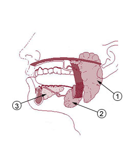 gland and secrete mucous into saliva to increase moisture for assistance in swallowing. The sublingual glands account for around 5% of saliva proction. The third and final pair of salivary glands are the submandibular glands which are located inferior to the mandibular ramus and inferior as well as posterior to the sublingual glands. The submandibular secrete serous as well as mucous secretions. Serous secretions contain amylase for starch breakdown. Despite being around half the weight of the parotid glands, submandibular glands account for around 75% of saliva production. As well as the 3 main salivary glands, several hundred minor salivary glands are present, mainly located in the palatial and buccal regions.
1. Parotid Gland 2. Submandibular gland 3. Sublingual gland. Image courtesy of Wikipedia under commons licence
Pharynx
- Once swallowing has occurred, food continues down the pharynx, commonly known as the throat. The Pharynx is involved as a pathway for both food and air, so when swallowing occurs, the epiglottis covers the trachea to avoid choking as a reflex. The length of the pharyx is about ~13cm in an adult and consists of 3 parts: nasopharynx (most superior origin), oropharynx and laryngopharynx (most inferior). Food descends from the oropharynx to the laryngopharynx. The pharynx extends from the underside of the skull to the cricoid cartilage level anteriorly, and to the sixth cervical vertebra posteriorly. Food then enters the oesophagus.
Oesophagus
- The oesophagus lies posterior to, and is continuous to the laryngopharynx. It essentially connects the pharynx (superiorly) to the stomach (inferiorly) of the GI tract. Peristalsis occurs initially in the oesophagus. This is a process by which food is pushed along the GI tract using circular smooth muscles behind the bolus to allow one way movement of food and the use of longitudinal smooth muscle to push the bolus forward towards the stomach. The process of peristalsis in the oesophagus usually takes ~9s. Presuming a bolus gets stuck during the first peristaltic wave, a second wave triggered by specific stretch receptors in the oesophagus around the bolus, will attempt to push the food down towards the stomach.
Stomach
- Inferior and continuous of the oesophagus lays the stomach (see diagram below). Here food is further broken down using stomach smooth muscles to increase food surface area for digestion. Furthermore, the stomach secretes hydrochloric acid which stands at a very low pH (~pH1-2) to inhibit bacterial action by preventing them from surviving at such a low pH. The stomach acts as storage of food for constant release rate into the small intestine.
- The 4 main sections of the stomach are the cardia, fundus, body and pylorus (in the order food travels through). The fundus contains fundic glands where chief cells are situated. Chief cells secrete pepsinogen which converts food proteins to peptides through digestion. Parietal cells, present in the fundic, cardia and pyloric regions of the stomach secrete gastric acid which contains mainly hydrochloric acid to lower the pH. The main function of the pyloric region is to use the muscles of the stomach to push food and chyme into the small intestine at a constant and manageable rate via the pyloric sphincter which allows unidirectional movement.

Diagram of location of stomach in relation to Oesophagus and small intestine. Image courtesy of Wikipedia under commons licence